Figure 2.
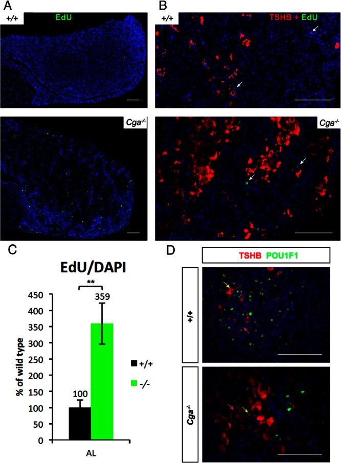
Thyrotrope hypertrophy and hyperplasia are associated with proliferation of TSHB-negative cells. A, Pituitaries were stained for EdU, which marks proliferating cells in S phase with green and counterstained with DAPI that makes the nuclei blue. B, Pituitary sections stained for TSHB in red, EdU in green, and nuclei in blue showed no costaining of TSHB and EdU in either genotype. Arrows indicate EdU-positive cells. Clusters of thyrotropes are interspersed with TSHB-negative cells. Scale bar, 100 μm (A, B, and D). Mice were 8 weeks of age. C, The proportion of DAPI positive cells that are EdU positive is increased in the Cga−/− mice. Ratio is compared with wild type. D, Thyrotropes stained with TSHB (red) from representative areas of 8-week-old pituitaries show a mix of POU1F1 positive (green) and negative cells in both genotypes. Nuclei were counterstained with DAPI.

