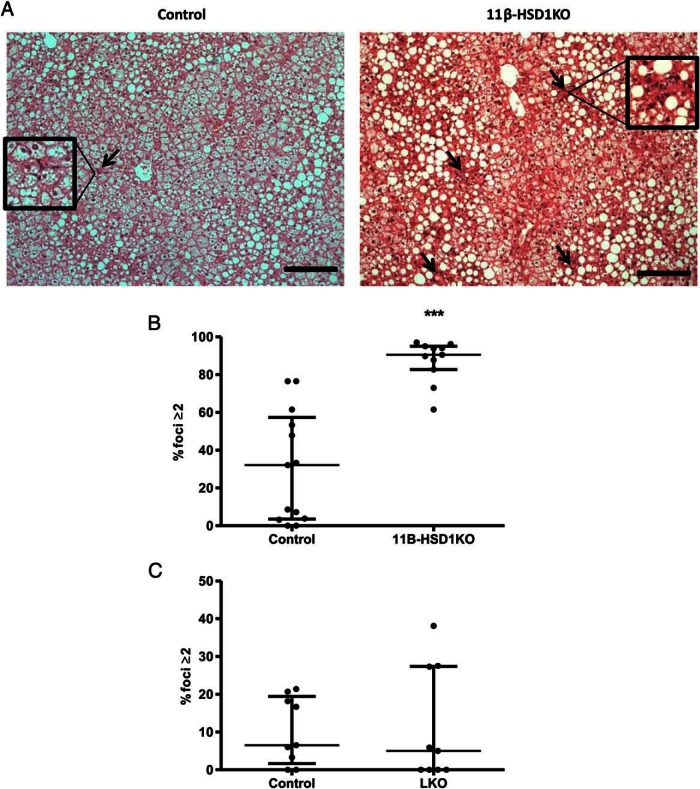
Figure 4.
Loss of 11β-HSD1 results in an increased frequency of hepatic inflammatory foci compared with control after 16 weeks of ALIOS diet. A, H&E-stained sections cut at 5 μm show inflammatory foci (arrows) in livers of controls and 11β-HSD1KO mice, areas inside boxes highlight inflammatory foci (magnified an additional ×2). B, Dot plot showing % fields of view with 2 or more inflammatory foci; using the Mann-Whitney test, there was a significant increase in 11β-HSD1KO mice compared with controls, which was not seen in livers of LKO mice when compared with their controls (C). ***, P < .001, interquartile range; n = 13 (control), n = 11 (11β-HSD1KO), n = 9 (LKO control), and n = 9 (LKO). Foci per field of view at ×200 magnification. Scale bars, 200 μm.

