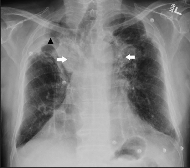
Figure 1.

A 71-year-old man with a history of chronic obstructive pulmonary disease, pulmonary hypertension, and prior pulmonary tuberculosis infection presenting with progressive dyspnea, diagnosed with tuberculosis-associated fibrosing mediastinitis. Chest posteroanterior radiograph showing bilateral peri-hilar soft tissue densities (white arrows) with right apical, pleural thickening and volume loss (black arrow head).
