Figure 5.
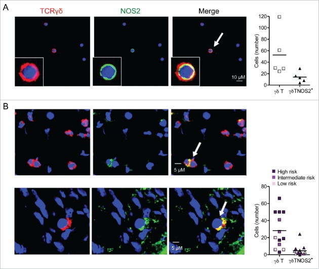
γδ Tcells infiltrating mouse and human melanoma are able to express NOS2. (A) Representative confocal microscopy images showing γδ T cells positive for NOS2 from cells derived from TdLNs of Ret mice and stained with antibodies to TCR γδ (red), NOS2 (green) and counterstained with DAPI (blue). Bars 10 µM. 40 X objective (left). Quantification of γδ T cells and γδ T cells positive for NOS2 from 500 to 1,500 cells (right). Experiments were performed five times. (B) Representative digital microscopy images of 7 µm sections from frozen biopsies of human primary melanoma (patient 14 and 12, respectively). Sections were stained with antibodies to TCR γδ (red) and NOS2 (green). Nuclei were counterstained with DAPI (blue). Bars 5 µM. 40 X objective (left). Quantification of γδ T cells and γδ T cells positive for NOS2 in 13 patients with detected γδ T cells (right). Patients were divided into three groups with low, intermediate or high risk of disease's relapse according to Breslow thickness. Arrows indicate NOS2-expressing γδ T cells.

