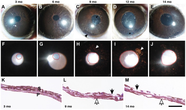
Fig. 1.
YBR mice exhibit a form of pigment-dispersing iris disease. YBR mice exhibit a progressive iris disease with age, characterized by iris stromal atrophy and pigment dispersion. (A-E) The top row of panels shows broad-beam illumination of YBR eyes at each of the indicated ages to assess the presence of dispersed pigment within the anterior chamber and iris stromal morphology. Mo, months. (F-J) The bottom row of panels shows transillumination to assess the degree of iris depigmentation, as revealed by areas where reflected light passes through the iris (white arrows). Pupils are outlined by red dotted lines. With transillumination, reflected light passes through the iris, after reflecting off the posterior pole of the eye. Normally, this reflected light would be blocked by the iris. (A,F) Three-month-old YBR eyes have normal iris morphology with clearly evident iris details. (B,G) By 6 months, mutant mice exhibited mild iris disease characterized by the mild accumulation of iris pigment in the peripupillary region. (C,H) By 9 months, YBR eyes exhibited iris stromal atrophy characterized by thinning of the iris stroma and peripupillary atrophy (black arrow head, C) and mild transillumination defects (white arrow, H). The degree of iris atrophy progressively worsened with age. Accumulation of dispersed pigment was greatest in the inferior angle (black arrow, C) owing to gravity. (D,I) At 12 months, some of the YBR eyes exhibited vascularization of the cornea (black asterisk, D), and most eyes showed worsening transillumination defects (white arrow, I). (K) In a young YBR mouse, the iris was well developed and had an intact stroma (S) and pigment epithelium (P). (L) The iris of an old YBR mouse exhibited moderately atrophied stroma (black arrow) and intact pigment epithelium (open arrow). (M) In severe cases, the stroma was largely atrophied with areas where it was essentially non-existent (black arrow). The iris pigment epithelium of these mice remained remarkably intact considering the overall condition of the iris (open arrows). Scale bar: 50 µm.

