Fig. 3.
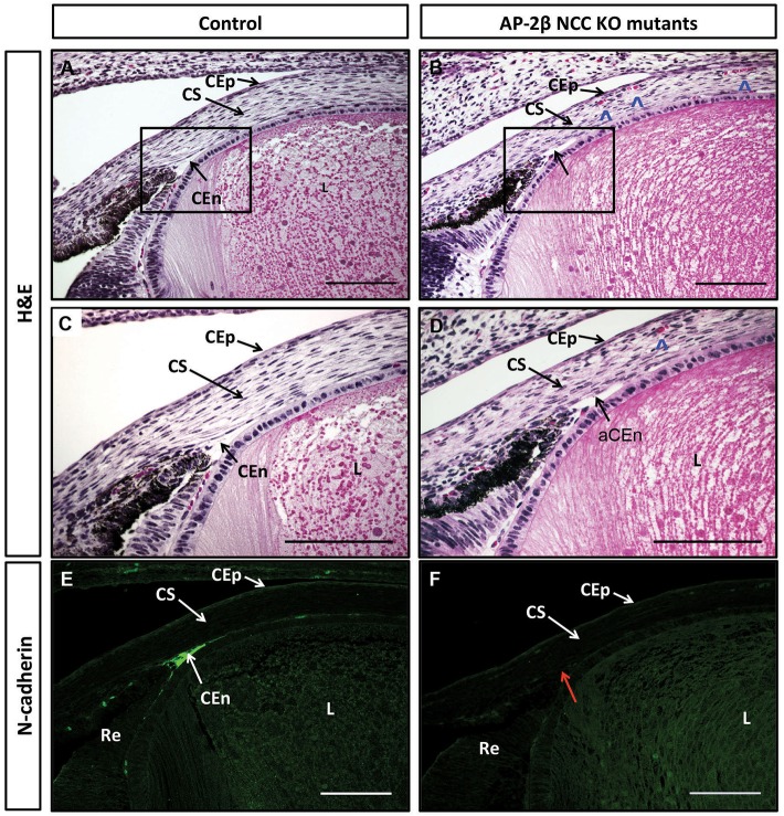
Embryonic corneal defects in E18.5 AP-2β NCC KO mutant embryos. Coronal sections of E18.5 anterior segments stained using either H&E (A-D) or immunofluorescence for N-cadherin expression (E,F) for controls (A,C,E) or AP-2β NCC KO mutants (B,D,F). Boxed regions in A and B are shown in greater detail in C and D. Red arrow in F shows that corneal endothelium does not stain for endothelial cell marker N-cadherin in absence of Tfap2b. Blue arrowheads in B and D indicate presence of red blood cells in mutant corneal stroma. CEn, corneal endothelium; aCEn, absent corneal endothelium; CEp, corneal epithelium; CS, corneal stroma; L, lens; Re, retina. Scale bars: 100 μm.

