Fig. 8.

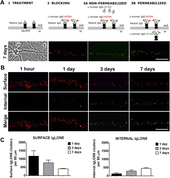
IgLON5 antibodies produce internalization of IgLON5 clusters. a Panel 1: Hippocampal neurons treated for 7 days with IgG-positive IgLON5 antibodies. Panel 2: IgG bound to IgLON5 on the dendrite surface labeled live with an excess of anti-human IgG Alexa Fluor 594 (red). Panel 3A: The saturation of the neuronal surface prevents that the anti-human IgG Alexa Fluor 488 (green) attaches to the surface. Panel 3B: After the neuron is permeabilized and incubated with the anti-human IgG Alexa Fluor 488, the green fluorescence localizes the human IgG attached to internalized IgLON5 clusters. Scale bar = 5 μm. b The internalization of IgLON5 clusters is a time-dependent effect and parallels a decrease of IgLON5 clusters on the surface of the dendrite. c Quantification of the number of IgLON5 clusters remaining on the cell surface and internalized after the treatment
