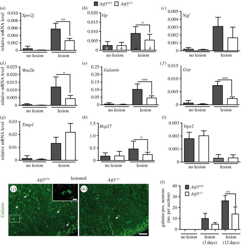
Figure 7.
ATF3 regulates injury-related expression of neuropeptides and other genes. (a–i) Unlesioned and injured FN of Atf3+/+ and Atf3−/− mice were subjected to qPCR analysis after 3 days of injury to quantify mRNA abundance of primers indicated. In wt mice (grey bars), facial nerve injury resulted in induction of Sprr2j (a), Vip (b), Ngf (c), Wnt2b (d), Galanin (e), Grp (f), Timp1 (g) and the known ATF3 target gene Hsp27 (h). In contrast to wt mice, induction of Sprr2j, Vip, Ngf, Wnt2b, Galanin, Grp and Hsp27 mRNA abundance was reduced upon facial nerve lesion in Atf3 mutant mice (white bars). Timp1 mRNA induction was more pronounced upon ATF3 loss-of-function (g). The Vip2 receptor (Vipr2) was downregulated by facial nerve injury in wt and ATF3-deficient mice (i). Numbers in bars in (b) reflect independent biological replicates for experiments in (a–i). (j–l) Confirmation of reduced galanin expression in ATF3-deficient mice upon facial nerve injury. Deafferented wt (j) and ATF3-deficient (k) FN were stained with anti-galanin directed antibodies. In wt mice (j), galanin localized in secretory vesicle-like structures (see inset) of FMNs (some labelled with an arrow). The number of galanin immunoreactive FMNs was reduced in Atf3 mutant mice (k). (l) Quantification of galanin positive neurons without and 3 and 12 days after lesion. Data are presented as mean ± s.d. **p ≤ 0.01. Scale bar (j,k) = 50 µm; inset = 5 µm.

