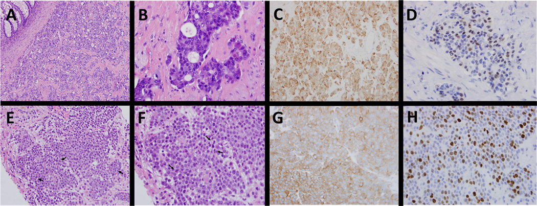
Figure 1.
Comparison of histologic features between primary rectal neuroendocrine tumor and liver metastasis. Histologic examination show a submucosal mass consisting of nested/organoid proliferation of variably small cells with stippled chromatin with some cells exhibiting pink granular cytoplasm (A-B). Immunohistochemical stains are diffusely positive for synaptophysin (C) and chromogranin (not shown), with variable CDX2 staining (D). Panels E-H highlight similar features from the liver biopsy with uniform cells (E-F), scattered mitotic figures (arrows), diffuse synaptophysin positivity (G) and Ki67 proliferative index 40–50% (H).

