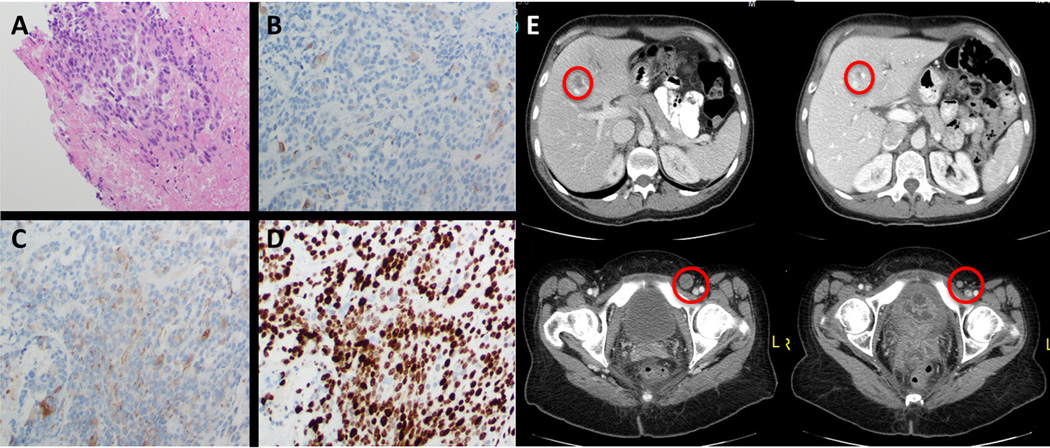
Figure 3.
Histologic features and radiographic response to vemurafenib and trametinib in BRAFV600E mutant high grade rectal neuroendocrine tumor from case 2. Microscopic examination demonstrates organization of variably small cells (A) staining weakly positive for CD56 (B) and chromogranin (C) with a Ki67 proliferative index of over 70% (D). Dramatic radiographic response to therapy is shown in panel E.

