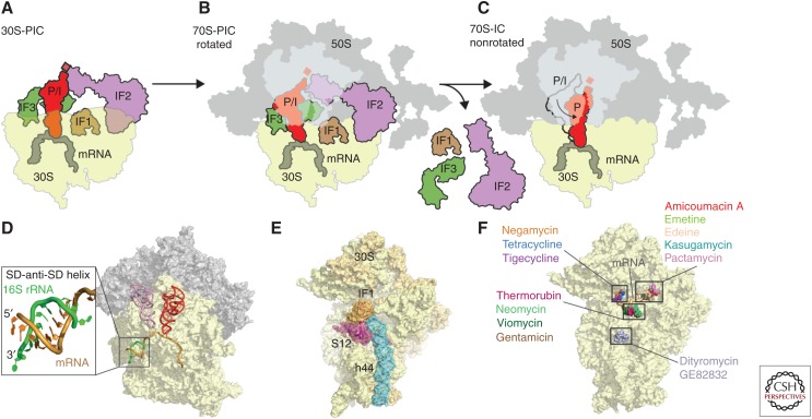
Figure 3.
Initiation of translation. Schematic assembly of the (A) 30S preinitiation complex (PIC), (B) 70S-PIC, and (C) 70S-IC during translation initiation with 30S subunit (yellow), 50S subunit (gray), messenger RNA (mRNA) (dark gray), initiator transfer RNA (tRNA) (red), initiation factors (IFs): IF1 (brown), IF2 (purple), and IF3 (green). (D) Crystal structure of the prokaryotic ribosome with zoom onto the interaction of the Shine–Dalgarno (SD) sequence of canonical mRNAs (orange) with the anti-SD at the 3′ end of the 16S ribosomal RNA (rRNA) (green), including P-site tRNA (red) and E-site tRNA (pink) (Yusupova et al. 2006). (E) Crystal structure of IF1 (brown) bound to the 30S subunit (yellow) with highlighted h44 (blue) of the 16S rRNA and ribosomal protein S12 (red) (Carter et al. 2001). (F) Binding sites of dityromycin (PDB 4NVU) (Bulkley et al. 2014), gentamicin (PDB 4V53) (Borovinskaya et al. 2007a), thermorubin (PDB 3UXT) (Bulkley et al. 2012), viomycin (PDB 3KNH) (Stanley et al. 2010), neomycin (PDB 2QAL) (Borovinskaya et al. 2007a), negamycin (PDB 4RBH) (Polikanov et al. 2014c), tetracycline (PDB 4G5K) (Jenner et al. 2013), tigecycline (PDB 4G5T) (Jenner et al. 2013), amicoumacin A (PDB 4RB5) (Polikanov et al. 2014a), edeine (PDB 1I95) (Pioletti et al. 2001), kasugamycin (PDB 1VS5) (Schuwirth et al. 2006), pactamycin (PDB 4RBB) (Polikanov et al. 2014c), and emetine (PDB 3J7A) (Wong et al. 2014), on the small subunit (SSU) (yellow). P/I, Peptidyl/initiation.

