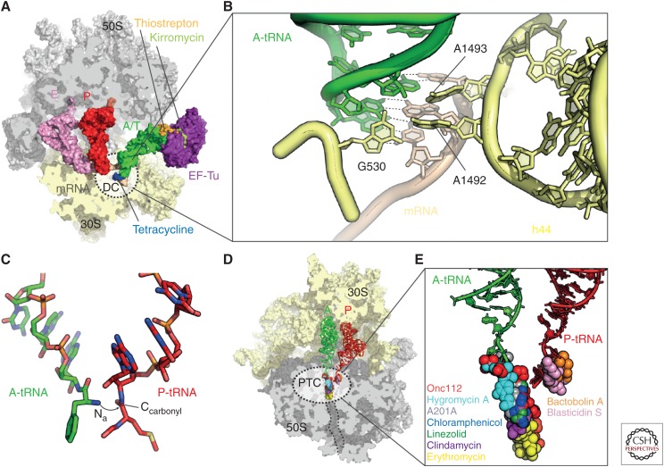
Figure 4.
Decoding and peptide bond formation on the ribosome. (A) Crystal structure of the ribosome in complex with elongation factor Tu (EF-Tu) (purple), A/T-transfer RNA (tRNA) (green), P-tRNA (red), E-tRNA (pink), kirromycin (light green), messenger RNA (mRNA) (brown), 30S subunit (yellow), and 50S subunit (gray) (Voorhees et al. 2010) with superimposed binding positions of thiostrepton (orange) (Harms et al. 2008) and tetracycline (blue) (Jenner et al. 2013). The decoding center (DC) on the 30S subunit is depicted as dashed-lined sphere. (B) DC within the small ribosomal subunit with A-tRNA (green), mRNA (brown), and 16S ribosomal RNA (rRNA) nucleotides G530, A1492, and A1493 (yellow) (Demeshkina et al. 2012). (C) Positions of the A-tRNA (green) and P-tRNA (red) within the peptidyltransferase center (PTC) of the 50S subunit. The nucleophilic attack of the A-tRNA α-amino group (Nα) onto the P-tRNA carbonyl-carbon (Ccarbonyl) is indicated with an arrow. (D) Overview and (E) zoom onto the binding sites of Onc112 (PDB 4ZER) (Seefeldt et al. 2015), hygromycin A (PDB 4Z3R) (Polikanov et al. 2015), A201A (PDB 4Z3S) (Polikanov et al. 2015), chloramphenicol (PDB 3OFC) (Dunkle et al. 2010), linezolid (PDB 3DLL) (Wilson et al. 2008), clindamycin (PDB 3OFZ) (Dunkle et al. 2010), erythromycin (PDB 3OFR) (Dunkle et al. 2010), bactobolin A (PDB 4WWE) (Amunts et al. 2015), and blasticidin S (PDB 4L6J) (Svidritskiy et al. 2013) within the PTC (dashed lines in D) of the 50S subunit (gray) with A-tRNA (green) and P-tRNA (red).

