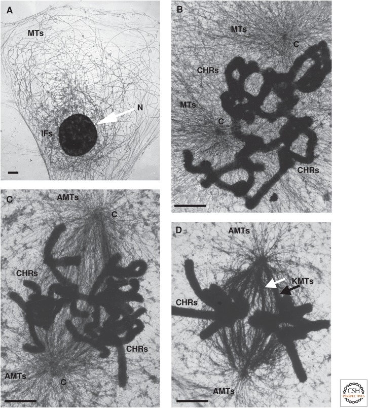
Figure 2.
Electron micrographs of cells as they form a mitotic spindle. Rat kangaroo PtK1 cells were cultured on gold grids coated with a thin layer of plastic and carbon, lysed with 0.2% Triton X100 and 1 mm MgCl2, in a PIPES-HEPES buffer, pH 7.2, and then fixed with 2% paraformaldehyde and 0.1% glutaraldehyde. These samples were quenched in 0.2 mg/mL NaBH4 in 1:1 ethanol:phosphate-buffered saline, then stained with a monoclonal antibody against tubulin, followed by a rabbit antimouse IgG bound to 10-nm colloidal gold, followed by fixation in osmium tetroxide, and then by drying with the critical-point method. Microtubules (MTs) in the interphase and mitotic cells are nicely contrasted, and the chromosomes are stained by osmium. (A) Interphase: The nucleus (N) contains decondensed chromatin. Intermediate filaments (IFs) surround the nucleus. (B) Early prometaphase: CHRs, chromosomes; C, centrosome. (C) Late prometaphase: AMTs, astral microtubules. (D) Metaphase: KMTs, kinetochore microtubules. Scale bars, 1 µm. (Images kindly provided by Mary Morphew, University of Colorado, Boulder.)

