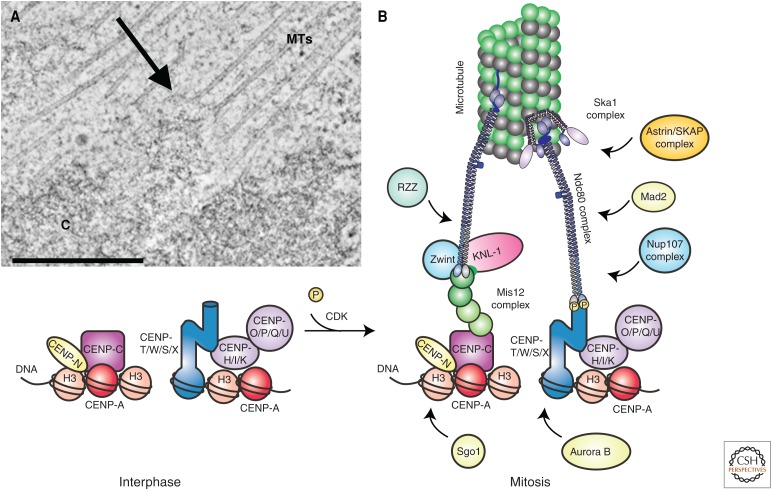
Figure 4.
Structure of the kinetochore. (A) Slice from an electron tomogram of a rat kangaroo PtK1 cell in prometaphase. A chromosome (C) and the associated microtubules (MTs) are easy to distinguish in the tomogram; their point of connection is the kinetochore (arrow). (B) Diagram of kinetochore composition and structure. At mitotic entry, phosphorylation (P) by activated CDK–cyclin-B promotes assembly of the outer kinetochore on a platform of constitutive kinetochore proteins. For information about the kinetochore proteins shown here, see Cheeseman 2014. CDK, cyclin-dependent kinase. (Reprinted, with permission, from Cheeseman 2014, © Cold Spring Harbor Laboratory Press.)

