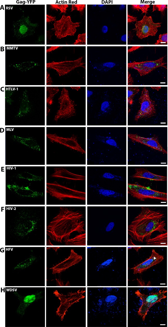
FIG 1.
Cellular localization of transient Gag-YFP expression in HeLa cells. HeLa cells were transiently cotransfected with 4:1 weight ratios of untagged Gag to YFP-tagged Gag in addition to homologous envelope plasmids as described in Materials and Methods. (A) Rous sarcoma virus (RSV); (B) mouse mammary tumor virus (MMTV); (C) human T-cell leukemia virus type 1 (HTLV-1); (D) murine leukemia virus (MLV); (E) HIV-1; (F) HIV-2; (G) human foamy virus (HFV); (H) walleye dermal sarcoma virus (WDSV). Optical sections close to the bottom of the cells were standardly used for collecting representative images of Gag-YFP localization. Nuclei were identified with DAPI stain (blue), and actin filaments were identified with the ActinRed 555 ReadyProbes reagent (red). Gag-YFP expression was identified by green fluorescence. Scale bar, 10 μm.

