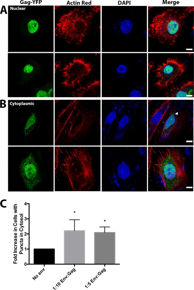
FIG 2.
Differential walleye dermal sarcoma virus (WDSV) Gag-YFP localization by WDSV Env expression. HeLa cells were transiently transfected as described in Materials and Methods with 1:4 weight ratios of WDSV Gag-YFP and untagged WDSV Gag in addition to WDSV Env expression plasmids at ratios of either 1:10 or 1:5. Cells were fixed, DAPI stained, and stained with ActinRed 16 h posttransfection. Images collected by confocal microscopy were used to evaluate cells for subcellular localization of WDSV Gag-YFP. (A) Representative examples of HeLa cells with no WDSV Gag-YPF puncta observed in the cytosol from transfections with no Env. (B) Representative examples of HeLa cells with WDSV Gag puncta observed in the cytosol from 1:10 or 1:5 cotransfections. Scale bar, 10 μm. (C) Fold increase in percentage of cells with Gag in the cytoplasm in different Env cotransfection ratios based on analysis of 50 cells (n = 3).

