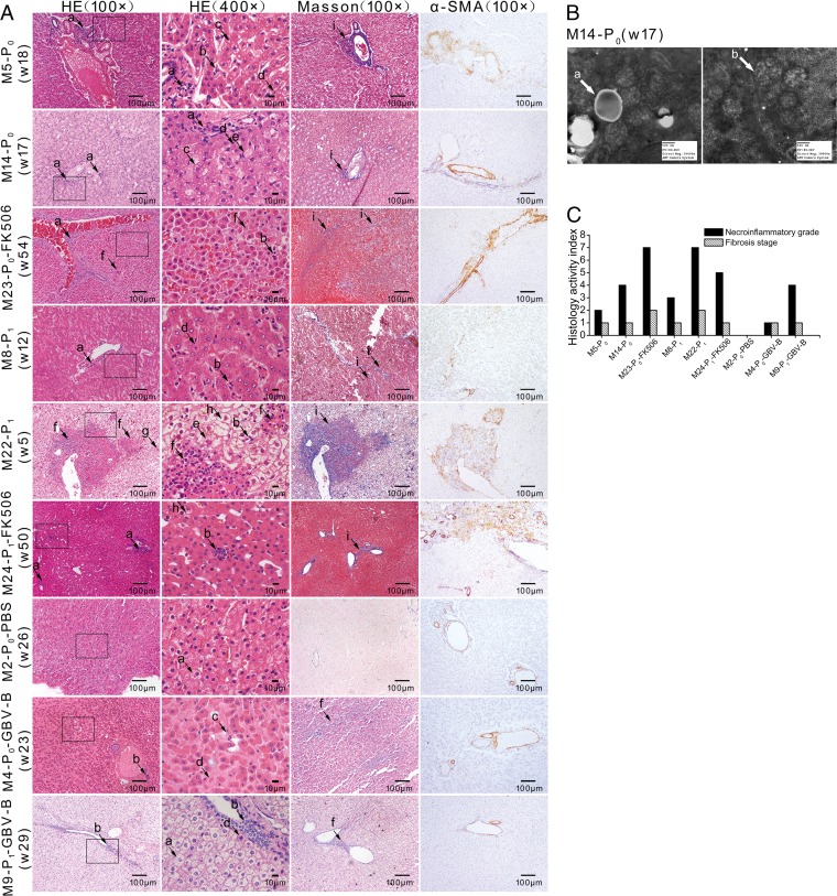
FIG 6.
Histopathological observation of liver tissues from infected marmosets. (A) Liver tissues of marmosets were stained with H&E, Masson's trichrome, and α-SMA. The lowercase letters with arrows indicate histopathological features: lymphocytic infiltrates (a), focal necrosis (b), steatosis (c), eosinophilic cells (d), ground glass liver cells (e), piecemeal necrosis (f), Kupffer cell enlargement (g), ballooning degeneration (h), and fibrous expansion (i). (B) Electron microscope image of marmoset M14-P0. The lowercase letters with arrows indicate histopathological changes: lipid droplets (a) and mitochondria (b). (C) Necroinflammatory grade and fibrosis stage were scored by the modified HAI system, in which necrosis and inflammation grades were on a scale of 0 to 18 and fibrosis was scored as 0 to 6.

