
FIG 3.
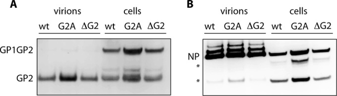
Immunoblot analysis of purified virions and infected cells. Vero cells were infected with wild-type, G2A-GPC, or ΔG2-GPC rCan virus (MOI of 0.1) for 48 h, and virions were purified from cell culture supernatants by centrifugation through 20% sucrose solution. Both the purified virions and infected cell monolayers were solubilized and subjected to SDS-PAGE and immunoblot analysis using GP2-directed MAbs G3 and G5 (39) (A) or NP-directed MAb AG12 (38) (B). The higher-molecular-size NP species may represent phosphorylated isoforms (73); the absence of these species in infected cell lysates could reflect errant phosphatase activity. The lower-molecular-size NP bands (*) are common degradation products. None of the identified species is present in lysates of uninfected cells. This experiment is representative of four independent virus growths, and average GPC and NP compositions are shown in Table 1.