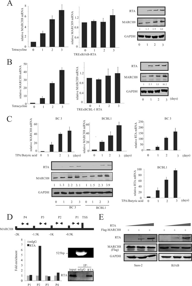
FIG 4.
RTA upregulated MARCH8. (A) TRExBJAB-RTA cells were induced with tetracycline for 0, 1, 2, and 3 days. RNA was isolated, and RT-PCR was performed. The relative levels of MARCH8 and MARCH9 were normalized to that of GAPDH (left and middle panels). Western blot analysis was used to detect the RTA and MARCH8 expression (right panel). Values represent the intensities of proteins normalized to that of GAPDH and compared to the signal obtained at day 0. (B) TRExBCBL1-RTA cells were induced by tetracycline for the indicated times. RNA was isolated, and RT-PCR was performed. The relative levels of MARCH8 and MARCH9 were normalized to that of GAPDH (left and middle panels). Western blot analysis was used to detect the RTA and MARCH8 expression levels (right panel). Values represent the intensities of MARCH8 proteins and were normalized against that of GAPDH and compared to the signal obtained at day 0. (C) BC3 and BCBL1 cells were reactivated using chemical inducer TPA and butyric acid for 0, 1, 2, and 3 days. RNA was isolated, and RT-PCR was performed. The relative levels of MARCH8 and RTA were normalized to that of GAPDH (top and lower right panels). Western blot analysis was used to detect RTA and MARCH8 expression (bottom panel). Values which represent the intensities of MARCH8 proteins were normalized against that of GAPDH and compared to the signal obtained at day 0. (D) RTA binds to the MARCH8 promoter. The schematic at the top shows the region of the MARCH8 promoter and the specific region targeted by the designed primers. Fifty million BCBL1 cells were treated with TPA and butyric acid for 24 h. Cells were harvested, and ChIP assays were performed as described in Materials and Methods using mouse IgG (mIgG) and anti-RTA antibodies; the cells were subjected to RT-PCR analysis using the primers described in Materials and Methods (P1 to P4). The DNA fragments after sonication and the immunoprecipitated RTA are shown to the right of the bar graph. 1K represents 1,000. (E) SAOS-2 and BJAB cells were cotransfected with 0, 10, and 20 μg RTA and 10 μg Flag-MARCH8. At 48 h posttransfection, the cells were lysed and subjected to WB analysis with RTA, Flag, and GAPDH antibodies.

