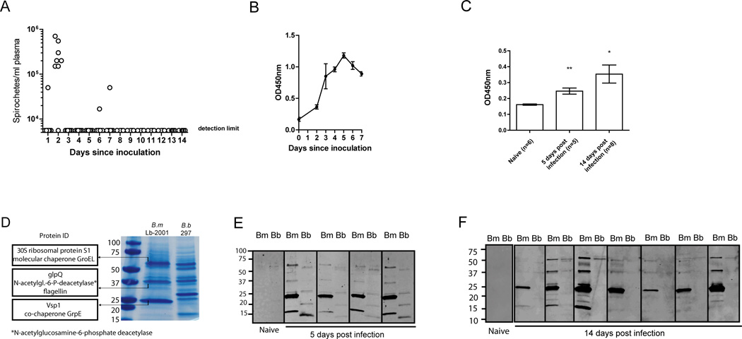
FIGURE 1. Borrelia miyamotoi infection induces a robust antibody response against a 23 kDa protein.
(A) Dark-field microscopy on plasma from eight C3H/HeN mice after intraperitoneal (i.p.) inoculation with B. miyamotoi LB2001 spirochetes. Horizontal lines represent mean concentrations, negatives represent the detection limit. (B) ELISA detecting IgM against B. miyamotoi LB-2001 whole-cell lysate. (C) IgG response to infection demonstrated by ELISA using sera from mice infected 5 or 14 days post-infection. (D) SDS-PAGE of B. miyamotoi whole-cell lysate stained by Coomassie brilliant blue, revealing abundant protein bands at ~23 kDa, ~39 kDa and ~60 kDa. These bands were analyzed by LC–MS/MS and the most abundant proteins are depicted on the left. For comparison, B. burgdorferi lysate (B.b 297) was run in the right lane. (E, F) Western blot analysis of nitrocellulose strips loaded with B. miyamotoi LB-2001 (Bm) and B. burgdorferi 297 (Bb) whole-cell lysates, detecting IgM in sera of four mice 5 days after B. miyamotoi infection (E) or IgG 14 days after infection in seven mice (F). Protein size in kDa is depicted on the left. * p<0.05, ** p<0.01. Error bars illustrate mean ± S.E.M.

