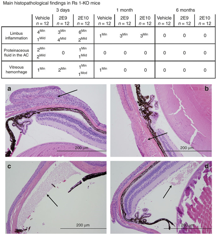
Figure 4.

Main histopathological findings in Rs1-KO mice intravitreally injected with AAV8-scRS/IRBPhRS at 2E9 and 2E10 vg/eye and vehicle. At 3 days after injection, a variable number of inflammatory cells were detected at the limbus of the injected eye of Rs1-KO mice treated with vehicle and with both 2E9 and 2E10 (a, arrow) vg/eye doses of AAV8-scRS/IRBPhRS. At 6 months, the limbal inflammation completely resolved in all groups, including the 2E10 vg/eye dose group (b, arrow). Some animals from each treatment group showed proteinaceous fluid deposits in the anterior chamber (c, arrow), and hemorrhages in the vitreous chamber, mainly at 3 days after injection. (d, arrow). All the described findings were attributed to the injection procedure. The inflammatory cells were counted and graded as minimal (<25), mild (25–50), moderate (50–75) and severe (>75). Hemorrhage was classified according to the number of red blood cells as minimal (<50), mild (50–100), moderate (100–150) and severe (>150). Presence of proteinaceous fluid was assessed and quantified as minimal, mild, moderate and severe. Hematoxylin and eosin; Bar = 200 µm (a–d). AC, anterior chamber.
