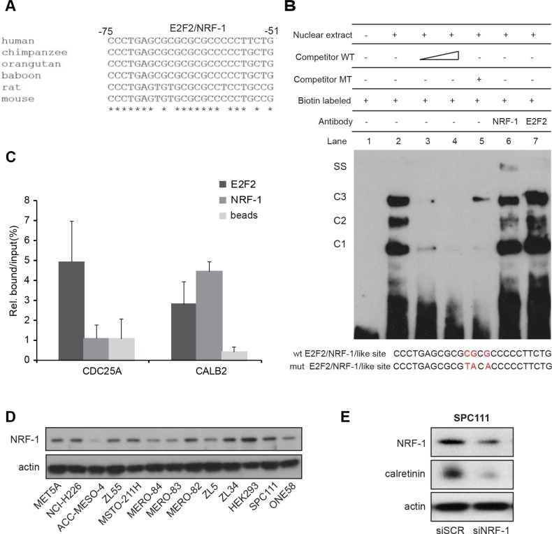
Figure 5. NRF-1 and E2F2 bind to the CALB2 promoter.
(A) Nucleotide sequence comparison reveals high homology in the CALB2 promoter regions of different species in the stretch containing E2F2/NRF-1 predicted binding sites. The analysis was performed using the ClustalW2 software. Asterisks indicate conserved nucleotides. (B) EMSA assay showing that a 25bp-oligo containing E2F2/NRF-1-like sites forms three DNA-protein complexes (C1, C2, C3) when incubated in with the ZL55 cells-derived nuclear extracts. Addition of a NRF-1 antibody but not of one against E2F2 resulted in a supershift (SS). (C) qPCR analysis on CDC25A and CALB2 promoter regions after ChIP-experiments. Data of two independent experiments were normalized to input. (D) NRF-1 protein expression in different cell lines. Actin is used as loading control. (E) NRF-1 silencing resulted in downregulation of calretinin expression.

