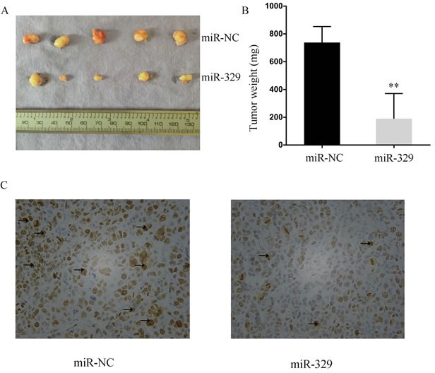
Figure 3. MiR-329 attenuated tumor growth in vivo.

A. The size of the xenograft tumor harvested from nude mice was evaluated. B. The weight of the xenograft tumor was shown by bar graph. C. Representative photographs of immunohistochemical analysis of Ki-67 expression in the xenograft tumor collected from nude mice. The positive staining was delineated by the arrow. Standard deviation of the mean was plotted for bar charts. (** P < 0.01)
