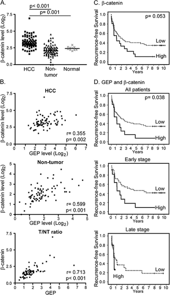
Figure 4. Clinical significance of GEP and β-catenin in HCC clinical specimens.

A. β-catenin transcript level was significantly up-regulated in HCC tumor (HCC, n = 77) compared with the paralleled tumor-adjacent non-tumor liver tissues (non-tumor, n = 77) and normal livers from healthy individuals (normal, n = 10). The lines indicate the median values. B. Expression levels of GEP significantly correlated with that of β-catenin in HCC tumor tissues (HCC), and in the paralleled non-tumor tissues. The tumor / non-tumor (T/NT) ratio showed the same trend. C. Kaplan–Meier recurrence-free survival plot according to β-catenin levels (log-rank test, p = 0.053). There were 56 patients with low β-catenin expression and 21 patients with high β-catenin expression (median recurrence-free survival of 24.5 months and 12.8 months, respectively). D. Patients (n = 77) were segregated into the low expression group (either one or both low in GEP and β-catenin) and the high expression group (both high in GEP and β-catenin). There were 61 patients in the low expression group (median recurrence-free survival, 24.5 months) and 16 patients in the high expression group (median recurrence-free survival, 12.6 months). Patients with high GEP and β-catenin levels were found to have poor recurrence-free survival (log-rank test, p = 0.038). When the patients were segregated into early and late tumor stages, patients with high GEP and β-catenin levels also demonstrated poor recurrence-free survival (log-rank test, p = 0.022).
