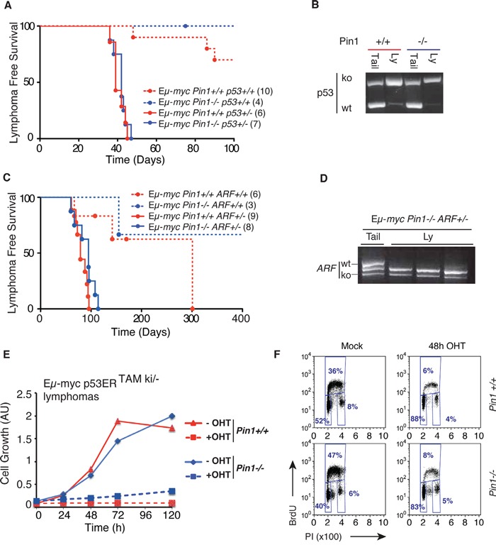
Figure 5. Delayed lymphomagenesis in the Pin1−/− background requires Arf/p53 activity.

A. Lymphoma-free survival in cohorts of Eμ-myc or control mice of the indicated Pin1 and p53 genotypes. The median survival was 39 days for Eμ-myc Pin1+/+p53+/− and 42 days for Eμ-myc Pin1−/−p53+/− mice (P=0.5959, Log-Rank, Mantel-Cox). Numbers within brackets indicate sizes of each cohort. B. Lymphomas arising in Eμ-mycp53+/− mice show p53 loss of heterozygosis (LOH) in either Pin1+/+ or Pin1−/− background, as shown by RT-PCR. One example of each is shown. Three Eμ-myc p53+/−Pin1−/− tumors were analyzed with the same outcome. C. Same as A. with the indicated Pin1 and ARF genotypes. Calculated Median survival was 79 days for Eμ-myc Pin1+/+ARF+/− and 95 days for Eμ-myc Pin1−/−ARF+/− mice (P=0.0959, Log-Rank, Mantel-Cox). Note that in both A. and C., Eμ-myc Pin1−/− mice with functional p53 and Arf show delayed lymphomagenesis relative to their Pin1+/+ counterparts, validating the results shown in Figure 1A. D. Lymphomas arising in Eμ-mycPin1−/−ARF+/− mice show ARF loss of heterozygosis (LOH). Three tumors and one tail from Eμ-myc Pin1−/−ARF+/− mice were analyzed by RT-PCR. E. Growth curves of Eμ-myc p53ERTAMki/− lymphomas overexpressing the anti-apoptotic protein Bcl2. Cells were cultured either in the presence (100 nM) or absence (Mock) of OHT. Cell growth was assessed with the Cell Titer Glo assay. F. Cell cycle analysis of Bcl2-expressing Eμ-myc p53ERTAMki/− lymphomas 48 hours after OHT administration.
