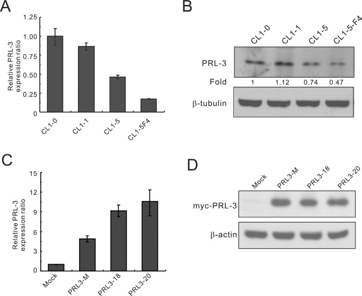
Figure 1. PRL-3 expression in lung cancer cell lines with increasing invasiveness and transfectants.
The invasive ability of the cell lines is as follows: CL1-0 < CL1-1 < CL1-5 < CL1-5F4. (A) PRL-3 mRNA expression levels in cell lines, as measured by real-time RT-PCR. (B) PRL-3 protein levels, as detected by Western blot analysis. β-tubulin was used as an internal control. (C) PRL-3 mRNA expression in the transfectants, as measured by real-time RT-PCR. CL1-5 cells were transfected with pCMV-Tag3B-PRL-3 or vector alone to establish stable cell clones, including mock, mixed, and single cell clones. TBP was used as an internal control. (D) PRL-3 protein expression in transfectants, as measured by Western blot analysis. β-tubulin was used as a loading control. Real-time RT-PCR was assessed in triplicate.

