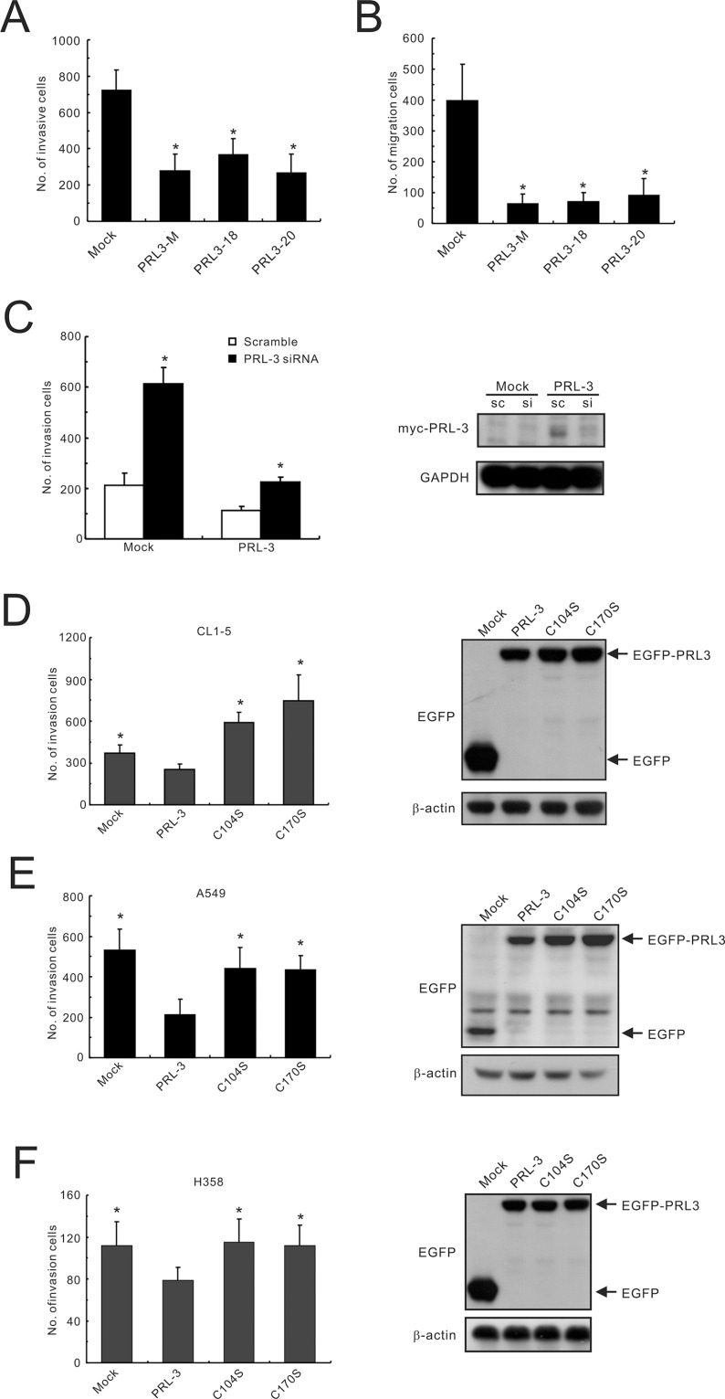
Figure 3. Inhibition of lung cancer cell invasion and migration by PRL-3 expression.
(A) Invasiveness and (B) migration ability of PRL-3 transfectants, as determined by a transwell apparatus. Mixed: mixed cell clones; Single: single cell clones PRL3-18 and PRL3-20); Mock; vector alone. The data are presented as the mean ± S.D. of three experiments. P values are compared with mock. (C) Invasive ability of lung cancer cells with PRL-3 knockdown. PRL-3 was silenced by a small-interfering RNA (siRNA) in PRL-3-overexpressing CL1-5 cells (PRL-3) and control cells (Mock), and then subjected to an in vitro cell invasion assay. Right panel: PRL-3 expression level after silencing. The data are expressed as the mean ± S.D. of three experiments, and P values are compared with scrambled siRNA control cells. (D–F) The effect of transient expression of wild-type PRL-3 and mutants PRL-3/C104S and PRL-3/C170S on cell invasive properties. The tested lung cancer cell lines include CL1-5 (D), A549 (E), and H358 (F). The ectopic expression levels of PRL-3 protein are displayed in the right panels, as detected by Western blotting. The data are presented as the mean ± S.D. of three experiments. *P < 0.05, compared with wild-type PRL-3 cells.

