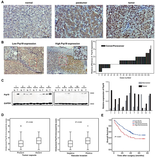
Figure 1. Prp19 overexpression in HCC indicates invasiveness and poor prognosis of HCC.

A. Representative images of IHC staining with Prp19 in liver tissues (from left to right: normal liver tissue, paratumor tissue and matched tumor tissue; Scale bar: 100μm). B. Representative view of Prp19 in HCC tumor tissues in IHC staining (Scale bar: 100μm). Micrograph indicated the amplified view of HCC tissue (original magnification ×400). Difference of Prp19 immunoactivity score in paired HCC tissue samples (N=169, right panel). C. Prp19 expression in tumor tissue (T) and paired paratumor tissue (N) from HCC patient specimens using western blot (left panel). Prp19 bands were quantified and shown in the bar chart after normalization (right panel). D. Relative immunoactivity score of Prp19 in HCC with or without tumor capsule (left panel) or vascular invasion (right panel). E. The overall survival rate between the low and high Prp19 expression groups in 169 HCC patients.
