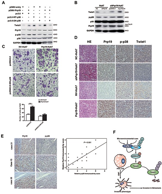
Figure 6. Prp19 mediates invasion of HCC via p38 MAPK/Twist1 pathway.

A. Huh7 cells were transfected with plasmids as indicated, cell lysates were analysed by immunoblot with indicated antibodies. B. shPrp19-Huh7 cells were transfected with myc-Prp19 and empty vector, followed by western blot. C. Prp19-Huh7 cells and NV-Huh7 cells were transfected with pcDNA3.0 or pcDNA3.0-DN-p38. The invasive capacities were analysed via Matrigel invasion chamber assay. D. Representative images of IHC staining of Prp19, p-p38 and Twist1 in the HCC tissues of orthotopic implantation model in nude mice (Original magnification ×200). E. Representative images of IHC staining with antibodies against Prp19, p-p38 in human HCC specimens (Original magnification ×200; left panel). Expression correlation of Prp19 and p-p38 was analysed in 80 HCC patients using IHC (right panel). F. Schematic presentation of the mechanism elucidating Prp19-mediated HCC invasion. NC, negative control; NV, null vector.
