
Figure 6. miR-101 targeting ZFX attenuates MAPK/Erk and Smad signaling.
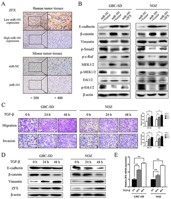
A. ZFX expression was detected by IHC in human and mouse tumor tissues. B. E-cadherin, β-catenin, vimentin, p-Smad2, p-c-Raf, MEK1/2, p-MEK1/2, Erk1/2, and p-Erk1/2 expression was analyzed by western blot analysis in cells transfected with an miR-NC/empty vector, an miR-101/empty vector, or an miR-101/ZFX plasmid. β-actin was used as a loading control. C. Representative figures and data from transwell assays conducted using GBC cells treated with 10 ng/mL TGF-β for 0, 24, and 48 h. D. Western blot analysis of E-cadherin, β-catenin, vimentin, and ZFX expression in GBC cells following treatment with TGF-β for 0, 24, and 48 h. E. Relative expression of ZFX mRNA in GBC cells treated with TGF-β for 0, 24, and 48 h.