
Figure 1. Lack of expression of MG53 in mouse brain tissue.
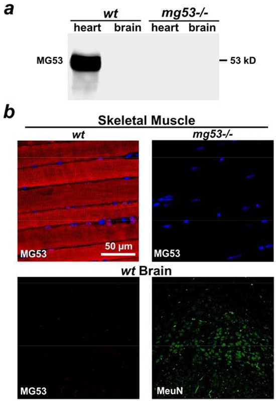
a. Western blotting showed that MG53 is present in the mouse heart, but not in the brain tissue. Tissues derived from mg53−/− mice were used as negative controls. b. Immunohistochemical staining revealed that MG53 is absent in brain tissue (lower left panel). Skeletal muscle tissue derived from wt mice (upper left panel) and mg53−/− mouse (upper right panel) were used as positive and negative controls respectively. NeuN staining was used to label neurons in brain slides (lower right panel).