
Figure 1. Hpa2 levels are decreased in bladder cancer.
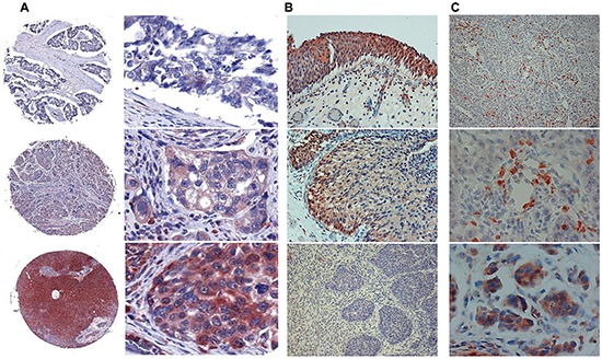
A. Bladder tissue array. Tissue array containing 69 biopsies of human bladder tumors was subjected to immunostaining applying anti-Hpa2 polyclonal antibody. Shown are representative photomicrographs of tumors that exhibit no or very weak staining (0-1; upper panels), moderate (+2, middle panels) or strong (+3; lower panels) staining. Original magnifications: left panels: x5, right panels: x40. B–C. Bladder biopsies. Bladder tumor biopsies were subjected to immunostaining applying anti-Hpa2 polyclonal antibody. Strong Hpa2 staining is detected in normal transitional epithelium of the bladder (B, upper panel) which is decreased substantially in bladder carcinomas (B, middle and lower panels). Original magnifications: x10. Hpa2 staining is also detected in immune cells within tumors (C, upper and middle panels), including macrophages giant cells (C, lower panel). Original magnifications: upper panel: x10, middle and lower panels: x40.