Figure 4.

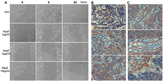
A. Hpa2 attenuates 5637 cell migration. Parental 5637 bladder carcinoma cells were plated in ibidi cell migration inserts apparatus (Planegg, Germany) until confluent. The barrier was then removed, cell cultures were washed and changed to serum-free medium, and migration into the defined cell-free gap was inspected in the absence (Con) or presence of the indicated concentration of purified Hpa2. Shown are representative photomicrographs taken before (0), 6, and 24 hours after the addition of Hpa2. Note that cell migration and wound closure is attenuated prominently by exogenous Hpa2. B. LOX staining. The bladder tissue array was subjected to immunostaining applying anti-LOX polyclonal antibody. Shown are representative photomicrographs of tumors that show weak (+1; upper panel), moderate (+2, middle panel) or strong (+3; lower panel) staining. C. LOX staining is also found enriched at apparently cell-cell borders (upper panel) and in endothelial (middle panel) and stromal cells (lower panel) within tumors. Original magnifications: x40.
