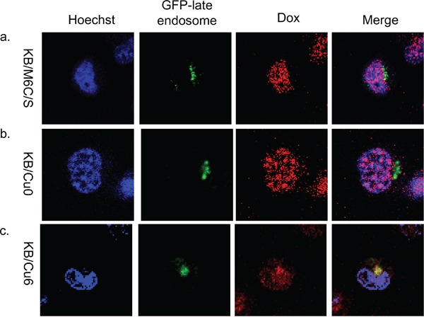
Figure 5. Subcellular localization of doxorubicin in mutant ATP7B expressing cells.

Subcellular localization of doxorubicin at 4 hours after doxorubicin exposure and washing with PBS in a. KB/M6C/S, b. KB/Cu0, and c. KB/Cu6 cells. GFP-late endosomes are green, doxorubicin is red. The nuclei are stained with Hoechst 33342 (blue).
