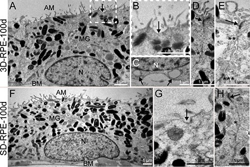
Figure 2. The ultrastructure of 3D-RPE and SD-RPE cells.

(A) 3D-RPE-100d cells had abundant apical microvilli (AM), melanin granules (MG) in the apical regions and the nucleus (N) in the basolateral regions. A basement membrane (BM) was visible. (B) A higher magnifications of the areas framed in Figure 2A showed caveolae (arrow) was distributed on the apical plasmalemma. (C) Caveolaes (arrow) were also distributed on the basolateral plasmalemma. (D) Tight junction (arrow) and gap junction (single asterisk) could be found. (E) Adherences junction (double asterisk) and desmosome (triple asterisk) could be found. (F) SD-RPE-100d cells had also abundant AM, MG in the apical regions and the nucleus (N) in the basolateral regions. A BM was visible. (G) Caveolae (arrow) was distributed on the apical plasmalemma in SD-RPE-100d cells. (H) Tight junction (arrow) and gap junction (single asterisk) could be found.
