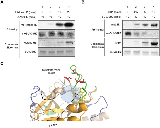
Figure 5. Automethylation of SUV39H2 and methylation of substrate proteins are exclusively correlated.

A-B. Recombinant SUV39H2 proteins were individually incubated with increasing amount of histone H3 protein (A) or LSD1 protein (B) in the presence of S-adenosyl-L-[methyl-3H]-methionine. The reaction mixtures were separated by SDS-PAGE, and the intensities of methylated proteins were detected by autoradiography, whereas the loading amounts of proteins were stained with Coomassie Brilliant Blue. C. The automethylation site (lysine 392) is located in a flexible loop (black dot line), which is suggested to form a part of the substrate lysine pocket. Pre-SET, SET and Post-SET domains are colored blue, orange, and green, respectively. SAM is shown as yellow stick and four cysteine residues corresponding to zinc binding are highlighted with red color.
