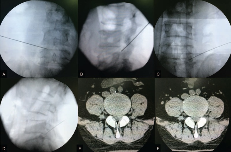
FIGURE 5.

Ideal position of needle by 1 puncture and successful removal of herniated disc. A = anteroposterior fluoroscopy at L4/5, B = lateral fluoroscopy at L4/5, C = anteroposterior fluoroscopy at L5/S1, D = lateral fluoroscopy at L5/S1, E = preoperative magnetic resonance imaging of lumbar disc herniation, F = postoperative magnetic resonance imaging confirming the removal of herniated disc.
