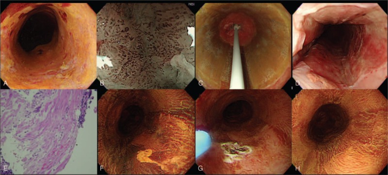
FIGURE 3.

Circumferential balloon-based radiofrequency ablation of early squamous neoplasia. (A) Lugol's staining showed a circumferential unstained lesion; (B) pretreatment evaluation with narrow band imaging and magnifying endoscopy to demonstrate the pattern of intra-epithelial papillary capillary loop (IPCL); (C) circumferential ablation catheter placed in the esophagus to ablate the lesion; (D) appearance of the mucosa after the second ablation; (E) the histology of endoscopic biopsy taken over the treatment area immediately after the RFA procedure, demonstrated the muscularis mucosa layer without viable tumor. (F) At 3 months, a small residual Lugol unstained lesion was noted and further eradicated with argon plasma coagulation (G); (H) 6 months after primary circumferential RFA, Lugol's staining showed no evidence of residual squamous neoplasia. A biopsy also confirmed the complete response. RFA = radiofrequency ablation.
