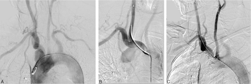
Figure 3.

Images of aneurysm. (A) Aortic arch aortography in a frontal view. This angiogram shows an aneurysm adjacent to the brachiocephalic trunk. (B) Selective brachiocephalic arteriogrphy before embolization. The aneurysm was fed by an abnormal bronchial artery branching from the brachiocephalic trunk. (C) Postprocedure angiogram in a frontal view. The ectopic bronchial aneurysm had been successfully embolized by interlocking coils. The right common carotid artery and subclavian artery were patent.
