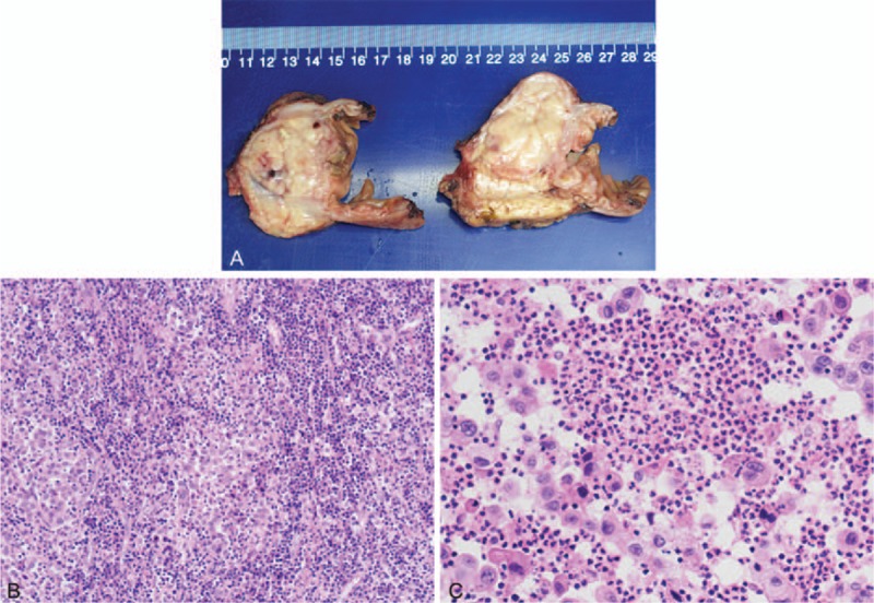
Figure 2.

(A) Pathological examination of the resected specimen revealed a mass (4.5 × 2.9 × 4.0 cm) originating from pancreatic head and was later diagnosed as undifferentiated adenocarcinoma. (B) A large number of lymphocytes infiltrated the tumor diffusely (Hematoxylin and eosin, 100 times). (C) Infiltration of polymorphonuclear leukocytes (PMNs) were visible within the tumor (Hematoxylin and eosin, 400 times).
