
Figure 1.
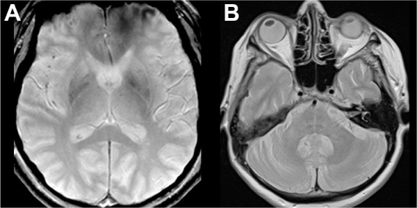
Qualitative radiological signs typically associated with MSA.
Notes: (A) Signal changes visible on T2*-weighted sequence in the lateral putamen. The patient was diagnosed as MSA-C. (B) Signal changes occurring in a cruciate pattern in the pons (“hot cross bun” sign) seen on proton density-weighted sequence. The patient was diagnosed as MSA-P.
Abbreviations: MSA, multiple system atrophy; MSA-C, multiple system atrophy, cerebellar subtype; MSA-P, multiple system atrophy, parkinsonian subtype.