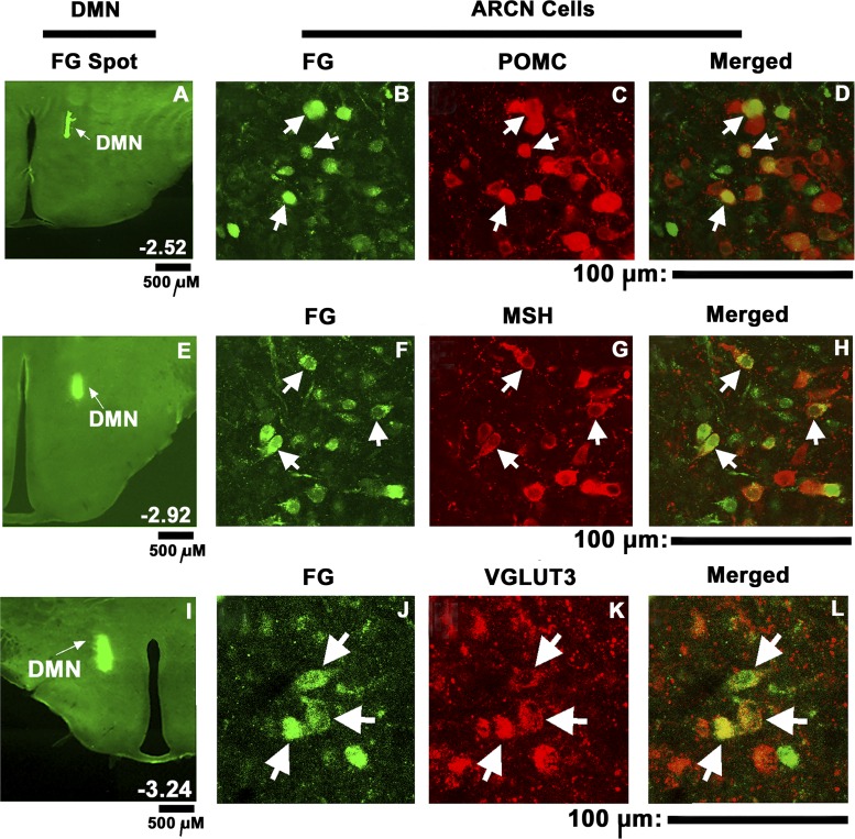
Fig. 6.
Retrograde tracing of ARCN projections to the DMN and immunohistochemistry. Fluoro-Gold microinjection site in the DMN (A, E, I). Subsequently, retrogradely labeled cells were observed in ipsilateral (B, F, J) and contralateral (not shown) ARCN. Immunohistochemistry showed cells staining for POMC (C), α-MSH (G), and VGLUT3 (K) in the ipsilateral ARCN. Merger of images in B and C showed that some ARCN cells retrogradely labeled with FG contained POMC (D, arrows). Merged images of F and G showed that some ARCN cells retrogradely labeled with FG contained α-MSH (H; arrows). Merger of images in J and K showed that some ARCN cells retrogradely labeled with FG contained VGLUT3 (L; arrows). Images of sections shown in A–D, E–H, and I–L are from different rats. FG, Fluoro-Gold; MSH, α-melanocyte stimulating hormone; POMC, proopiomelanocortin; VGLUT3, vesicular glutamate transporter-3. A, E and I: scale bars = 500 μm each; for other panels: scale bars = 100 μm each.

