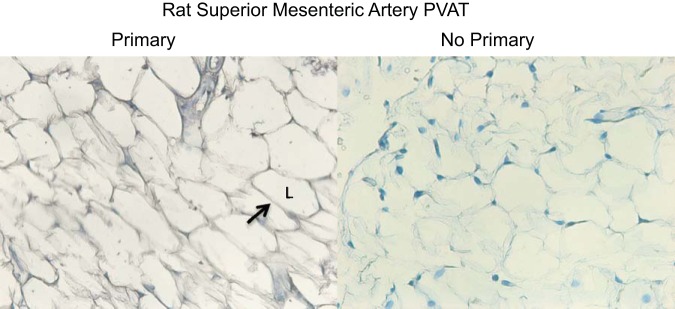
Fig. 1.
Immunohistochemical localization of chemerin to perivascular adipose tissue (PVAT) of rat superior mesenteric artery in experiments in which primary antibody directed against chemerin was present (left) and experiments in which the primary antibody was absent (right). Arrow points to an area of adipose cell cytoplasm that stains positively. L, lipid droplet. Images are representative of 4 different animals.

