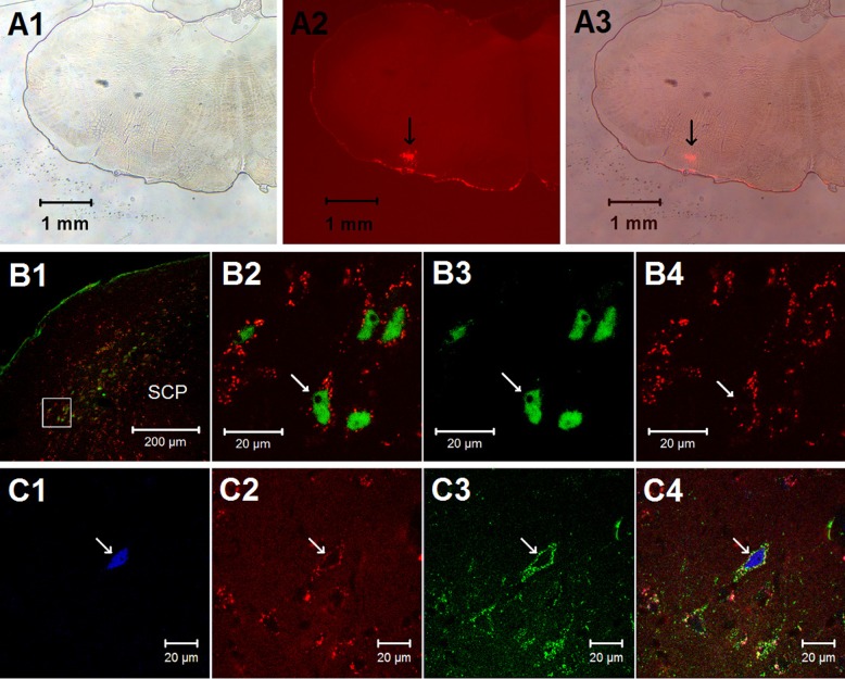
Fig. 1.
Histological staining in the external lateral parabrachial nucleus (elPBN) and the rostral ventrolateral medulla (rVLM). A1–A3: photomicrographs demonstrating a microinjection site of the retrograde microsphere tracer in the rVLM [bregma −12.12 mm; from the rat brain atlas of Paxinos and Watson (37)]. A1: bright-field image of a medulla section. A2: fluorescence image showing the microinjection site of the retrograde microsphere tracer in the medulla section. A3: merged image from A1 and A2. Arrows in A2 and A3 indicate the injection site located in the rVLM. B and C: confocal microscopic images showing double (B1–B4) and triple (C1–C4) fluorescent labeling in the elPBN (bregma −9.00 mm) of a rat following stimulation of cardiac sympathetic afferent nerves with epicardial application of α,β-methylene-ATP (α,β-meATP, 8 μg). B1: low-power photomicrograph. B2: higher magnification of the region within the box (located in the elPBN) in B1. B2 is a merged image from B3 and B4. Arrows in B2, B3, and B4 indicate a neuron labeled with c-Fos + tracer, c-Fos (green), and tracer (red), respectively. SCP, superior cerebellar peduncle. Arrows in C1, C2, C3, and C4 indicate a neuron containing c-Fos (blue), retrogradely transported microspheres originating from the rVLM (red), vesicular glutamate transporter 3 (green), and colocalization of the 3 labels.

