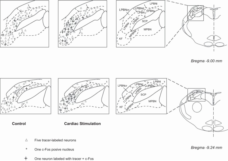
Fig. 2.
Distribution of c-Fos immunoreactivity and/or cells labeled with retrograde microsphere tracer in the elPBN following epicardial stimulation with α,β-meATP (8 μg) and in a sham-operated control. Two coronal sections of the pons ipsilateral to the tracer injection in the rVLM [atlas of Paxinos and Watson (37)] were selected from 1 animal in each experimental group. Each symbol represents a labeled cell(s). SCP, superior cerebellar peduncle; PBN, parabrachial nucleus; LPBN, lateral PBN; LPBNc, central part of LPBN; LPBNcr, crescent part of LPBN; LPBNd, dorsal part of LPBN; LPBNe, external part of LPBN; LPBNi, internal part of LPBN; LPBNv, ventral part of LPBN; MPBN, medial PBN; MPBNe, external part of MPBN; KF, Kölliker-Fuse nucleus.

