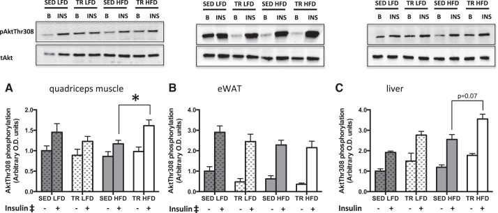
Fig. 5.
Insulin signaling is enhanced in skeletal muscle from previously trained mice fed a high-fat diet. Basal (gray bar) and insulin-stimulated (white bar) phosphorylation of Akt (∼60 kDa) at Thr-308 in quadriceps muscle (A), epididymal white adipose tissue (B), and liver tissue (C) of low-fat (LFD) or high-fat (HFD) fed sedentary (SED) or trained (TR) mice. Stippled bars indicate LFD to easily distinguish the two feeding treatments. Data are presented as means ± SE; n = 9. *Significantly different from SED HFD, P < 0.05. ‡Main effect of insulin, P < 0.05.

