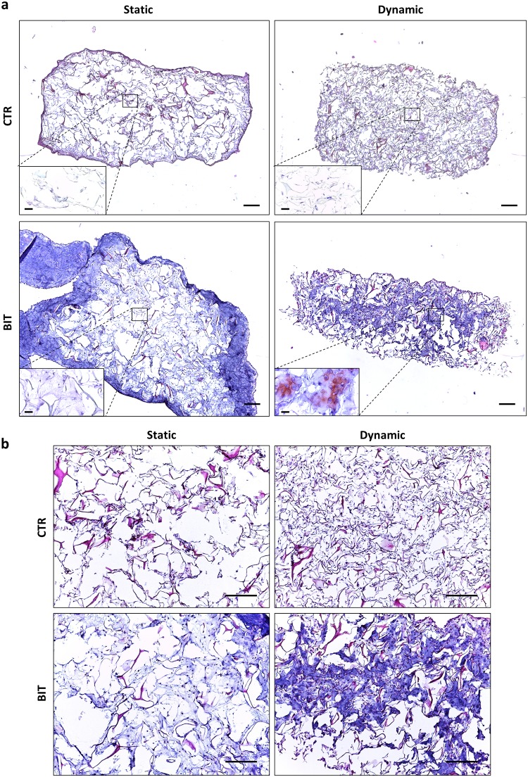
Fig 3. The combination of interstitial perfusion and BIT improves ECM neo-synthesis and distribution.
(a) Representative low-magnification pictures showing cell and ECM distribution in collagen sponges cultured for 21 days in static or dynamic conditions (program 1, n = 4) in the absence (CTR) or in the presence of BIT (HE staining, scale bars 100 μm). Inserts in the left low corner show the Safranin O staining in the scaffold core (scale bars 10 μm). (b) High-magnification pictures showing cell and matrix distribution in the scaffold core (HE staining, scale bars 100 μm).

