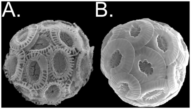
Fig 1. Representative images of E. huxleyi.

Scanning electron microscopy images of cells representative of (A) the lower calcifying strain CCMP371 and (B) the higher calcifying strain CCMP3266. The malformed-looking coccoliths seen in CCMP371 were commonly seen under all treatment conditions. Image sizes are not to scale.
