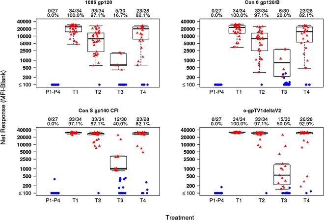
Fig 6. Peak IgG binding antibody response.
Binding magnitude of IgG responses to gp120 and gp140 antigens are shown as mean fluorescent intensity (MFI) in top, middle and lower panels, respectively. Positive responders are indicated in red and negative responders in blue. The mid-line of the box denotes the median and the ends of the box denote the 25th and 75th percentiles. The whiskers that extend from the top and bottom of the box extend to the most extreme data points that are no more than 1.5 times the interquartile range (i.e., height of the box) or if no value meets this criterion, to the data extremes. The number and percent positive responders in each group are shown above the graphs. Placebo recipients from all treatment groups are shown together. T1: MVA prime, sequential gp140 boost (M/M/P/P); T2 (MP/MP): concurrent MVA/gp140; T3 (D/D/M/M): DNA prime, sequential MVA boost; T4 (D/D/MP/MP): DNA prime, concurrent MVA/gp140 boost) or placebo.

