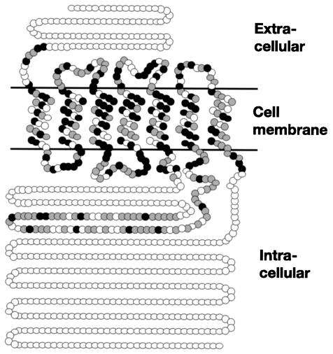
FIG. 1.
Predicted two-dimensional model of C. albicans Gpr1. Closed circles indicate identical amino acids, and shaded circles indicate conserved amino acids compared to S. cerevisiae Gpr1. The amino acid positions of the transmembrane regions are as follows: 100 to 122, 135 to 157, 177 to 199, 220 to 242, 264 to 286, 439 to 458, and 473 to 495. The secondary structure of Gpr1 was analyzed by using TMHMM (http://www.cbs.dtu.dk/services/TMHMM/).

